Patellar Tendinitis, also known as Jumper’s Knee, is a common condition that affects athletes and active individuals. It occurs when the patellar tendon, which connects the kneecap (patella) to the shinbone (tibia), becomes irritated or inflamed. This injury can lead to significant pain and limited mobility, making it difficult to perform physical activities.
Fortunately, advancements in medical imaging, particularly Musculoskeletal Ultrasound (MSK Ultrasound), have provided an innovative and non-invasive way to diagnose and treat patellar tendinitis. In this blog, we’ll explore how ultrasound technology is transforming the way this condition is managed, offering athletes and active individuals an effective path to recovery.
What is Patellar Tendinitis?
Patellar tendinitis is an overuse injury that causes inflammation of the patellar tendon, typically as a result of repetitive stress or sudden changes in physical activity. It’s most common in athletes who participate in sports that involve jumping, running, or repetitive knee movements.
Symptoms include:
- Pain in the front of the knee, just below the patella
- Swelling around the knee joint
- Stiffness or discomfort when moving the knee
- Difficulty with activities like running, jumping, or climbing stairs
Patellar tendinitis can affect individuals of all activity levels, not just athletes. However, it’s particularly common among active people such as runners, basketball players, and cyclists.
Why Choose Ultrasound for Patellar Tendinitis Diagnosis?
Benefits of MSK Ultrasound: Musculoskeletal ultrasound is an advanced imaging technique that uses high-frequency sound waves to create detailed, real-time images of soft tissues, including tendons and muscles. Unlike traditional imaging methods like X-rays or MRIs, ultrasound doesn’t require radiation or invasive procedures.
Comparison with Other Imaging Techniques: Traditional MRI scans and X-rays may not always provide the level of detail necessary to assess soft tissue injuries like patellar tendinitis. Ultrasound, however, allows for a dynamic view of the tendon, enabling doctors to observe the tendon’s movement, thickness, and condition. This real-time feedback helps in making a more accurate diagnosis.
How Does Patellar Tendinitis Ultrasound Work?
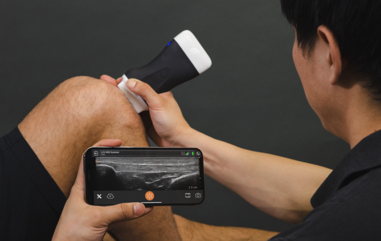
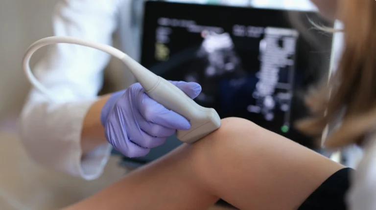
MSK ultrasound for patellar tendinitis involves applying a special gel to the skin near the knee, followed by a small handheld device called a transducer that sends sound waves into the body. These sound waves bounce off tissues and return to the transducer, creating a detailed image of the patellar tendon and surrounding structures.
Visualizing Tendon Damage: Ultrasound imaging allows doctors to assess the condition of the tendon, looking for signs of inflammation, tears, or changes in tendon thickness. This helps in determining the severity of the injury and crafting a personalized treatment plan.
Benefits of Patellar Tendinitis Ultrasound for Treatment
Accurate Diagnosis: The primary benefit of using Patellar Tendinitis Ultrasound is its ability to provide accurate, real-time imaging. By directly visualizing the tendon, ultrasound allows clinicians to determine the exact location and extent of the damage. This ensures that patients receive the right diagnosis and avoid the risks of misdiagnosis.
Guided Therapies: Ultrasound isn’t just useful for diagnosing patellar tendinitis; it also assists with treatments. For example, ultrasound guidance can improve the accuracy of injections (such as corticosteroid or PRP injections) by ensuring that the medication is delivered directly into the affected area. This results in more effective treatments and faster recovery times.
Reduced Recovery Time: Since ultrasound aids in the precision of treatments like physical therapy and injections, it helps reduce recovery time by ensuring the most appropriate therapy is applied immediately. This tailored approach ensures faster healing and better results.
Common Treatments for Patellar Tendinitis with Ultrasound Assistance
Physical Therapy: MSK ultrasound is often used alongside physical therapy to assess muscle function, tendon movement, and overall knee health. It allows physical therapists to track progress and adjust exercises based on real-time feedback from the ultrasound images.
Injection Therapy: In cases where inflammation is severe, ultrasound can guide injections of medications like corticosteroids or PRP (Platelet-Rich Plasma) directly into the inflamed tendon. This targeted approach helps reduce pain and inflammation more effectively than traditional methods.
What to Expect During a Patellar Tendinitis Ultrasound Exam
The ultrasound exam for patellar tendinitis is a quick, non-invasive procedure that typically lasts about 20-30 minutes. Here’s what to expect:
- Preparation: You may be asked to remove clothing from the lower half of your body and lie down with your knee exposed.
- Ultrasound Procedure: A gel will be applied to your knee, and the ultrasound technician will move a small transducer over the skin to capture images of the patellar tendon.
- Post-Procedure: There is no recovery time, and you can return to your daily activities immediately after the exam.
When to Seek Ultrasound Imaging for Patellar Tendinitis
If you’re experiencing symptoms of patellar tendinitis, such as knee pain, swelling, or limited mobility, seeking an ultrasound exam early on is crucial for effective treatment. Early diagnosis can help prevent the condition from worsening and ensure you start the right treatment sooner.
Indications for Ultrasound:
You should consider ultrasound imaging if:
- You’ve been experiencing knee pain that doesn’t improve with rest.
- You participate in activities that involve jumping or running.
- You have swelling or tenderness around your knee.
Why Choose MSK Ultrasound at OPTCI for Patellar Tendinitis?
At Optimal Performance & Therapy Centers of Indiana (OPTCI), we use the latest in Musculoskeletal Ultrasound technology to provide accurate diagnoses and effective treatments for patellar tendinitis. Our skilled physical therapists work closely with each patient, utilizing ultrasound to guide therapy and monitor healing progress.
We believe in offering a personalized treatment approach, which means that your rehabilitation plan is based on the results of your ultrasound exam. This ensures that you receive the most effective and targeted treatment for a quicker recovery.
Takeaway
Patellar tendinitis can be a painful and limiting condition, but thanks to advancements in ultrasound technology, diagnosis and treatment have never been more effective. If you’re struggling with knee pain or suspect you may have patellar tendinitis, consider scheduling an ultrasound exam at OPTCI. Our expert physical therapists are ready to help you recover and get back to doing what you love—faster and more efficiently.