Advanced imaging technologies have transformed the way specialists diagnose and manage complex vision conditions. Neuro-Ophthalmology Treatment focuses on disorders that affect the connection between the eyes and the brain. Because many of these conditions involve the optic nerve, brain pathways, or neurological structures, advanced imaging plays a crucial role in identifying the root cause of visual problems.
Modern diagnostic tools allow eye specialists to detect abnormalities earlier and develop more accurate treatment plans. Patients who seek Neuro-Ophthalmology Treatment from trusted providers such as Peregrine Eye and Laser Institute benefit from advanced imaging systems and experienced specialists who understand the complexity of neurological eye conditions.
Understanding Neuro-Ophthalmology
Neuro-ophthalmology is a specialized field that combines ophthalmology and neurology. It focuses on vision disorders that are related to the nervous system rather than the eye alone. Neuro-Ophthalmology Treatment addresses conditions that affect the optic nerve, visual pathways, and brain structures responsible for sight.
What Is Neuro-Ophthalmology?
Neuro-ophthalmology examines how the brain and eyes work together to create vision. When this connection is disrupted by disease, injury, or neurological conditions, patients may experience vision loss, double vision, or unusual visual symptoms. Neuro-Ophthalmology Treatment involves detailed diagnostic testing and specialized care to identify the underlying problem.
Highly trained specialists evaluate both neurological and ocular factors during Neuro-Ophthalmology Treatment. Clinics such as Peregrine Eye and Laser Institute provide comprehensive evaluations using advanced diagnostic technologies designed to detect even subtle abnormalities in the visual system.
Common Conditions Treated in Neuro-Ophthalmology
Several conditions may require Neuro-Ophthalmology Treatment, including disorders that affect the optic nerve and the brain’s visual pathways. These conditions often present symptoms that cannot be explained by routine eye examinations.
Common conditions include optic neuritis, papilledema, optic nerve tumors, double vision (diplopia), visual field loss, and neurological disorders that affect eye movement. Accurate diagnosis is essential because many of these conditions may be linked to serious medical issues such as brain tumors, autoimmune diseases, or increased intracranial pressure.
Specialists at Peregrine Eye and Laser Institute are experienced in evaluating these complex cases and providing advanced Neuro-Ophthalmology Treatment tailored to each patient’s needs.
The Role of Advanced Imaging in Neuro-Ophthalmology Treatment
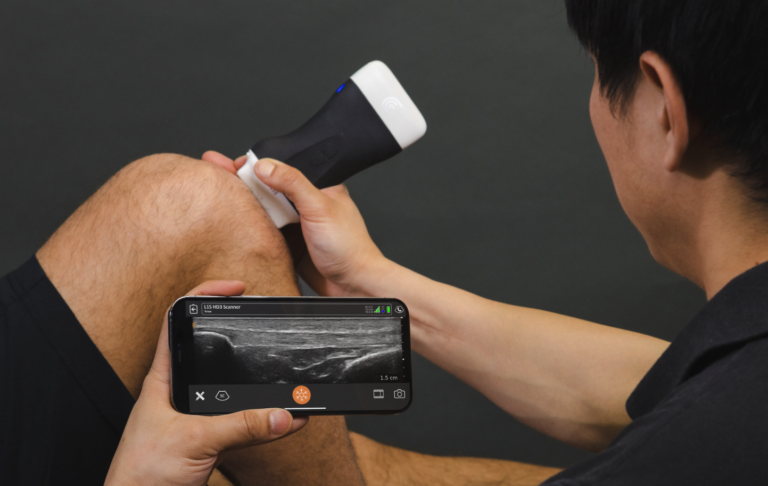
Advanced imaging technology is a vital component of effective Neuro-Ophthalmology Treatment. Many neurological vision problems cannot be diagnosed through basic eye exams alone. Imaging tools provide detailed views of the optic nerve, retina, brain structures, and visual pathways.
Why Imaging Is Essential for Accurate Diagnosis
Advanced imaging helps doctors detect abnormalities that may otherwise go unnoticed. Neuro-Ophthalmology Treatment often depends on imaging techniques that reveal inflammation, nerve damage, tumors, or structural changes in the brain.
These diagnostic tools allow specialists to identify the exact cause of symptoms such as unexplained vision loss, persistent headaches, or abnormal eye movements. Early and accurate detection is critical for preventing further damage to the visual system.
Patients seeking reliable Neuro-Ophthalmology Treatment often turn to Peregrine Eye and Laser Institute because of its advanced diagnostic capabilities and commitment to comprehensive patient care.
Benefits of Advanced Imaging Technology
Advanced imaging provides numerous benefits for both doctors and patients undergoing Neuro-Ophthalmology Treatment. These technologies allow for non-invasive testing, faster diagnosis, and more precise evaluation of neurological eye conditions.
High-resolution imaging also enables specialists to monitor disease progression and evaluate the effectiveness of treatment over time. This level of precision improves patient outcomes and helps prevent long-term vision complications.
Peregrine Eye and Laser Institute uses modern imaging systems that support accurate diagnosis and effective Neuro-Ophthalmology Treatment for a wide range of complex vision disorders.
Common Advanced Imaging Techniques Used in Neuro-Ophthalmology
Several advanced imaging technologies are commonly used in Neuro-Ophthalmology Treatment. Each method provides unique insights into the structure and function of the visual system.
Optical Coherence Tomography (OCT)
Optical Coherence Tomography is one of the most important imaging tools used in Neuro-Ophthalmology Treatment. This technology creates high-resolution cross-sectional images of the retina and optic nerve.
OCT helps specialists detect early signs of optic nerve damage and retinal changes that may indicate neurological disease. The detailed images produced by OCT assist doctors in diagnosing conditions such as optic neuritis and glaucoma-related nerve damage.
Magnetic Resonance Imaging (MRI)
Magnetic Resonance Imaging plays a critical role in Neuro-Ophthalmology Treatment because it provides detailed images of the brain and optic pathways. MRI scans can reveal tumors, inflammation, nerve compression, or other abnormalities affecting vision.
Specialists often recommend MRI when symptoms suggest that a neurological condition may be affecting the visual system. Early detection through MRI allows doctors to begin Neuro-Ophthalmology Treatment before significant vision loss occurs.
Computed Tomography (CT Scan)
CT scans are another useful imaging tool for Neuro-Ophthalmology Treatment. This technology uses X-rays to produce detailed images of bone structures and surrounding tissues.
CT imaging is particularly helpful when doctors suspect trauma, fractures, or structural abnormalities that may affect the optic nerve or eye socket.
Fundus Photography
Fundus photography captures detailed images of the retina and optic nerve. These images help specialists monitor changes in the optic nerve head and detect swelling or damage.
This imaging technique is frequently used during Neuro-Ophthalmology Treatment to track disease progression and evaluate treatment effectiveness.
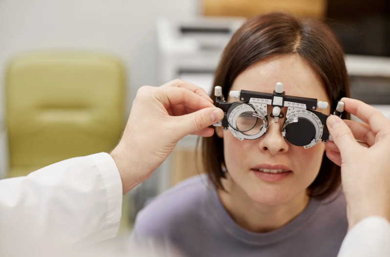
Visual Field Testing
Visual field testing measures a patient’s peripheral vision and detects blind spots or visual field loss. This test is essential in Neuro-Ophthalmology Treatment because it helps identify neurological conditions affecting the optic nerve or brain.
Visual field analysis provides valuable information that supports diagnosis and treatment planning.
How Advanced Imaging Improves Neuro-Ophthalmology Treatment
Advanced imaging has significantly improved the quality and accuracy of Neuro-Ophthalmology Treatment. These technologies allow doctors to detect problems earlier and develop personalized treatment plans.
Early Detection of Neurological Eye Diseases
Early detection is one of the most important benefits of advanced imaging in Neuro-Ophthalmology Treatment. Many neurological eye conditions progress slowly and may not show obvious symptoms at first.
Imaging technologies allow specialists to detect small changes in the optic nerve or brain structures before severe vision loss occurs.
More Accurate Treatment Planning
Detailed imaging results allow doctors to tailor Neuro-Ophthalmology Treatment to each patient’s specific condition. Accurate diagnosis leads to more effective treatment strategies and better outcomes.
Peregrine Eye and Laser Institute provides individualized care using advanced imaging technologies to guide every step of the treatment process.
Monitoring Treatment Progress
Advanced imaging also allows specialists to monitor the progress of Neuro-Ophthalmology Treatment over time. Regular imaging tests help doctors determine whether treatments are working and make adjustments when necessary.
This ongoing monitoring helps protect vision and improve long-term results for patients.
What Patients Can Expect During Neuro-Ophthalmology Imaging Tests
Most imaging procedures used in Neuro-Ophthalmology Treatment are non-invasive and comfortable for patients. Many tests require little or no preparation before the examination.
During imaging tests, patients may be asked to focus on a target, remain still while images are captured, or undergo scanning procedures such as MRI or OCT. These tests are typically quick and painless.
After the imaging process, specialists analyze the results and develop a personalized Neuro-Ophthalmology Treatment plan based on the findings. Clinics such as Peregrine Eye and Laser Institute ensure that patients fully understand their diagnosis and available treatment options.
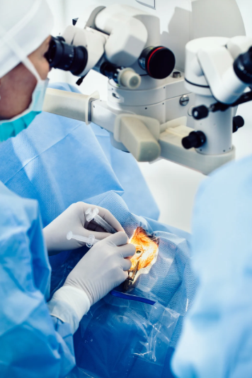
Why Choosing the Right Eye Specialist Matters
Complex neurological vision conditions require specialized expertise and advanced diagnostic equipment. Choosing the right provider for Neuro-Ophthalmology Treatment can make a significant difference in the accuracy of diagnosis and the success of treatment.
Peregrine Eye and Laser Institute is recognized for its commitment to high-quality eye care and advanced diagnostic technologies. The institute’s experienced specialists provide comprehensive Neuro-Ophthalmology Treatment designed to address the unique needs of each patient.
By combining expert medical knowledge with modern imaging tools, Peregrine Eye and Laser Institute offers patients reliable care for even the most challenging neuro-ophthalmic conditions.
Takeaway
Advanced imaging has become an essential component of modern Neuro-Ophthalmology Treatment. Detailed diagnostic tools allow specialists to identify complex neurological eye conditions with greater precision and begin treatment earlier. Accurate imaging improves diagnosis, supports effective treatment planning, and helps monitor patient progress over time.
Patients seeking reliable and comprehensive Neuro-Ophthalmology Treatment can trust Peregrine Eye and Laser Institute for expert care and advanced diagnostic technology. With experienced specialists and modern imaging systems, the institute provides high-quality treatment designed to protect vision and improve overall eye health.
Frequently Asked Questions (FAQ)
What is Neuro-Ophthalmology Treatment?
Neuro-Ophthalmology Treatment focuses on diagnosing and managing vision problems related to the brain, optic nerve, and nervous system. It addresses conditions that affect how the brain processes visual information.
Is advanced imaging painful?
Most imaging procedures used in Neuro-Ophthalmology Treatment are painless and non-invasive. Tests such as OCT, visual field testing, and fundus photography are quick and comfortable for patients.
How long do neuro-ophthalmology imaging tests take?
Many imaging tests used in Neuro-Ophthalmology Treatment take between 10 and 30 minutes. More detailed scans such as MRI may take longer depending on the condition being evaluated.
Who needs Neuro-Ophthalmology Treatment?
Patients who experience unexplained vision loss, double vision, abnormal eye movements, or neurological symptoms affecting sight may require Neuro-Ophthalmology Treatment. Specialists evaluate these symptoms using advanced imaging and diagnostic tools.
Can imaging detect brain-related vision problems?
Yes. Advanced imaging technologies such as MRI and OCT can detect structural changes in the brain and optic nerve that may cause vision problems. These tools play a crucial role in accurate Neuro-Ophthalmology Treatment.